Maitland's Vertebral Manipulation
Book information
Description
Front cover Half title page Dedication Maitland's Vertebral Manipulation Copyright page Table of Contents Contributors Biography Geoffrey Douglas Maitland MBE AUA FCSP FACP (Monograph), FACP (Specialist Manipulative Physiotherapist) MAppSc (Physiotherapy) Preface Acknowledgements In Memoriam: Kevin Banks (1959–2012) Glossary Chapter 1 The Maitland Concept: assessment, examination and treatment of movement impairments by passive movement Chapter 2 Clinical reasoning: from the Maitland Concept and beyond Chapter 3 Communication and the therapeutic relationship Chapter 4 Management of cervical spine disorders: a neuro-orthopaedic perspective Chapter 5 Management of thoracic spine disorders Chapter 6 Management of lumbar spine disorders Chapter 7 Management of sacroiliac and pelvic disorders Chapter 8 Sustaining movement capacity and performance Appendix 4 Recording References 1 The Maitland Concept: Chapter contents A personal commitment to the patient A mode of thinking: the primacy of clinical evidence Techniques Techniques of management Examination Palpatory techniques Assessment Analytical assessment Pretreatment assessment Assessment during every treatment session Proving the value of a technique Progressive assessment Retrospective assessment Final analytical assessment Conclusion References 2 Clinical reasoning: Chapter contents Introduction Clinical reasoning defined Clinical reasoning and evidence-based practice Critical thinking and clinical reasoning Importance of skilled clinical reasoning to expert practice Clinical reasoning and the bio-psychosocial model of health and disability Clinical reasoning as a hypothesis-oriented and collaborative process The physiotherapist’s thinking The patient’s thinking Health perspectives (pain, illness, self) Clinical reasoning as collaboration between therapist and patient Clinical reasoning and knowledge Clinical reasoning and cognition/metacognition Skilled questioning important to critical thinking and learning Socratic questioning Skilled questioning important to clinical practice Clarification for precision Clarification for accuracy Clarification for relevance Clarification for completeness Screening questions Effects of therapist’s questioning/manner on the patient Facilitating application of bio-psychosocial practice: clinical reasoning strategies and hypothesis categories Clinical reasoning strategies Diagnosis Management Hypothesis categories Activity capability/restriction Participation capability/restriction Patient perspectives on their experience Pathobiological mechanisms Physical impairments and associated structures/ tissue sources Contributing factors Precautions and contraindications to physical examination and treatment Management and treatment Prognosis Interpreting information across different hypothesis categories Pattern recognition Complexity of clinical reasoning Errors of clinical reasoning Improving clinical reasoning: learning through clinical reasoning Understanding clinical reasoning theory Facilitated clinical reasoning through case studies and real patients Self-reflection worksheets and clinical pattern diaries Mind maps Lateral/creative thinking We are all imposters References 3 Communication and the therapeutic relationship Chapter contents Introduction Therapeutic relationship The physiotherapist’s role in the therapeutic relationship Research and the therapeutic relationship The therapeutic relationship and physiotherapy education and practice Communication and interaction Aspects of communication Shaping of interactions Shaping of a therapeutic climate: listening and communication Communication techniques Paralleling Immediate-response questions Key words and phrases Bias Purpose of the questions and assuming Purpose of the questions Assuming Pain and activity levels The process of collaborative goal setting Critical phases of the therapeutic process Welcoming and information phase Subjective examination Planning of the physical examination Physical examination Ending a session Evaluation and reflection of the first session, including treatment planning Reassessments Retrospective assessment Final analytical assessment Verbatim examples Welcoming and information phase Initial assessment: subjective examination ‘First question’ – establishing main problem Behaviour of the symptoms History of the problem Initial assessment: physical examination Palpation Summarizing the first session: collaborative treatment planning and goal setting Directive interaction Collaborative goal setting by asking questions Beginning of a follow up session: subjective reassessment Reassessment Effects of self-management strategies Reassessments of physical examination tests During a treatment intervention Treatment and education of bodily awareness Directive communication Mirroring, guiding by asking questions, including reassessments Retrospective assessments (after three to five treatments) Final analytical assessment Conclusion References 4 Management of cervical spine disorders: Chapter contents Introduction Epidemiology of neck, head and facial pain Common syndromes of the cervical region and their presentations Whiplash-associated disorders (WAD) Headache Cervical nerve root lesion Clinical reasoning and the bio-psychosocial model A definition for pain Pain mechanisms Placing pain mechanisms into a reasoning framework Input dominant mechanisms Nociception Types of nociceptive neurons Location of nociceptive neurons Activation of nociceptive neurons Nociceptors and nociception Speed of messaging Transmission of messages via second order neurons Mechanical nociception Ischaemic nociception Inflammatory nociception Neurogenic contributions to inflammation Summary of clinical patterns from inflammation Clinical detection of nociceptive mechanisms Pain associated with changes in the nervous system Nerves aren’t normally that sensitive Injuries to peripheral nerves Blood flow Axoplasmic flow Clinical detection of peripheral neuropathic pain Centrally mediated mechanisms Central sensitization The brain and pain Brain changes in pain Mirror neurons and context change Output mechanisms Sympathetic nervous system Endocrine response Parasympathetic nervous system (PNS) The immune system Motor system Descending modulatory control Examination of the cervical region Subjective examination Planning the physical examination Physical examination Starting out the physical examination Observation Functional assessment Testing positions Ongoing analysis of your patient and reassessment Physical examination of the nervous system Palpation of peripheral nerves Response to nerve palpation Palpation related to peripheral neuropathic pain Palpation of the nerves of the head, neck and upper limb The trigeminal nerve (V) Palpation of the cervical nerve roots and the brachial plexus Neurodynamic testing Responses to neurodynamic testing Using structural differentiation in neurodynamics Neurodynamics relating to cervical conditions The nerve roots, trunks and cords of the brachial plexus The classical upper limb neurodynamic tests (ULNT) with structural differentiation Altering the test and/or start position Pre-cervical spine treatment screening – implications for examination Cervical arterial dysfunction (CAD) Craniovertebral instability Symptoms and signs of cervical instability Clinical testing of craniovertebral instability Examination of the cervical spine through mobilization techniques Passive mobilization Mobilization linked to context change The treatment of the cervical region Information and communication Shaping beliefs through pain education Passive mobilization techniques Specific mobilization treatments Selecting the correct technique The correct testing position Incorporating context change into treatment Manual therapy and central sensitization Manipulation Treatment with reference to neurodynamics Treating the containers Neural mobilization techniques Massage Self-treatment and management Treatment dose and ongoing intervention Graded exposure in order to progress treatment References 5 Management of thoracic spine disorders Chapter contents Introduction: thoracic spine and the Maitland Concept Clues in the subjective examination to thoracic spine involvement Symptoms at and around thoracic spine levels, and in areas neurally related to T1–12 Chronic conditions, which are not resolving with treatment Patient example Improvement of signs and symptoms in areas remote from the thoracic spine after passive mobilization of the mid-thoracic spine region Patient examples: manipulative procedures applied to the thoracic spine Mrs W Miss A Thoracic mobilization/manipulation: when to incorporate techniques to directly address neurodynamic signs/symptoms in the healing process of a lumbar disc lesion Analysis of role of detailed examination in determining when to use thoracic passive mobilization techniques and associated interventions Physical examination Observation Present pain Functional demonstration (and differentiation where appropriate) Brief appraisal Thoracic rotation Thoracic flexion, extension Flexion Extension Thoracic lateral flexion When applicable tests Combined movement tests Compression movement tests Tap test Slump test Palpation Areas of sweating and temperature changes Soft-tissue changes Bony changes and position tests Passive accessory intervertebral movements (PAIVMs) Differentiation test by palpation Passive range of physiological movements of single vertebral joints (PPIVMs) C7–T4 (flexion) Starting position Method C7–T4 (flexion/extension) Starting position Method C7–T4 (lateral flexion) Method C7–T4 (rotation) Method T4–11 (flexion/extension) Starting position Method T4–11 (lateral flexion) Starting position Method T4–11 (rotation) Starting position Method Examination and treatment techniques Mobilization Posteroanterior central vertebral pressure () Starting position Method Local variations Uses Rotary posteroanterior intervertebral pressures Starting position Method Uses Transverse vertebral pressure () Starting position Method Local variations Uses Posteroanterior unilateral vertebral pressure () Starting position Method Local variations Uses Posteroanterior unilateral costovertebral pressure () Starting position Method Local variations Uses Thoracic spine: rotation to the right (T2–12) Starting position Method Mobilization of the ribs (R2–12) Thoracic traction Upper thoracic spine (TT ) Starting position Method Lower thoracic spine (TT ) Starting position Method Local variations Precautions Uses Grade V manipulation overview Action (by the therapist to the patient) Mechanical response (occurring within the patient) Indications Precautions (Maitland 1986) Contraindications (Gibbons & Tehan 2001) Absolute contraindications Relative contraindications (at risk for adverse reactions) Principles and guidelines for manipulative thrust of the thoracic spine Intervertebral joints C7–T3 (lateral flexion ) Starting position Method Intervertebral joints T3–10 (PAs ) Starting position Method Intervertebral joints T3–10 (longitudinal movement ) Starting position Method Intervertebral joints T3–10 (rotation ) Starting position Method Additional examination and treatment techniques for thoracic disorders Cervical anteroposterior unilateral vertebral pressure () Starting position Method Local variations Precautions Uses Slump test PKB/Slump Starting position Method Straight leg raising (SLR) Starting position Method Anteroposterior sternochondral/costochondral joint mobilization Starting position Method References 6 Management of lumbar spine disorders Chapter contents Introduction Demedicalization and conceptualization of NSLBP Demedicalization Conceptualization Clinical assessment Treatment/advice to the patient Referral of patients with acute low back pain Scope of practice of physiotherapists regarding NSLBP Pillars of physiotherapy practice Paradigms International Federation of Orthopaedic Manipulative Physiotherapists’ competencies and scope of practice Treatment objectives Sense of control Optimizing movement capacity Psychosocial aspects in treatment Phases of NSLBP and physiotherapeutic treatment Classifications, subgroups and models Clinical reasoning Hypotheses generation and testing Experiential knowledge, clinical patterns Prognosis and clinical prediction rules Clinical prediction rules Reflective practice Examination of the lumbar spine: subjective examination Introduction to the assessment process Main problem Perceived disability Localization and quality of symptoms Behaviour of symptoms ‘Making features fit’ History of symptoms Recent history Previous history Medical and health screening questions Typical patterns of clinical presentation Physical examination Planning the physical examination procedures Precautions to examination procedures Physical examination and the lumbar spine Observation Functional demonstration Active tests lumbar spine Neurological conduction testing Neurodynamic testing Slump test Palpation Passive testing Passive physiological intervertebral movements (PPIVMs) Passive accessory intervertebral movements (PAIVMs) Examination of motor control impairment Mobilization and manipulation treatment techniques Accessory movements and variations Physiological movements and variations: mobilizations, manipulations Neurodynamic techniques Combination of arthrogenic techniques and neurodynamic mobilizations (example) Direct neurodynamic mobilization techniques Lumbar spine mobilization and manipulation techniques linked to clinical and supporting research evidence Integrated treatment Where there is evidence of neurogenic and myogenic impairments coexisting Where there is evidence of arthrogenic and neurogenic impairments coexisting Where there is evidence of arthrogenic and myogenic impairments coexisting Where there is evidence of arthrogenic, myogenic and neurogenic impairments coexisting Where there is protective muscle spasm coexisting with arthrogenic, other myogenic and neurogenic impairments Case studies References 7 Management of sacroiliac and pelvic disorders Chapter contents Introduction Applied theory and evidence supporting practice Form closure, force closure, mobility Local and global stabilizing muscle system The posterior oblique sling The deep longitudinal sling The anterior oblique sling Classification model Treatment Consideration of other factors leading to PGP Clinical reasoning Clinical reasoning and assessment procedures Evidence based practice Subjective examination Specific objectives of subjective examination Information phase Subjective examination 1. Main problem 2. Area of symptoms 3. Behaviour of symptoms and activity-levels 4. History (Hx) 5. Special questions (SQ) Planning of the physical examination (‘structured reflection’) Physical examination Observation Gait Posture Active movements of the trunk Forward bending Backward bending Side-bending Rotation Movements from below upwards Active movements of the hip Functional tests of load transfer Stork test Active straight leg raise test Pain provocation tests The posterior pelvic pain provocation test (P4 test; Ostgaard 2007) Distraction test (anterior distraction and posterior compression test) Compression test (anterior compression and posterior distraction; Fig. 7.24) Gaenslen’s test (Fig. 7.25) Sacral thrust test (Fig. 7.26) Patrick’s Faber test (Fig. 7.27) Long dorsal SI ligament test (Fig. 7.28; Vleeming et al. 1996, 2002) Palpation of the symphysis pubis (Fig. 7.29) Passive tests Positional tests Position of the innominates in supine (Figs 7.30 and 7.31) Position of the pubic tubercles Position of the innominates in prone (Fig. 7.32) Position of the sacrum in prone (Figs 7.33 and 7.34) Passive mobility tests Passive physiological movements of the innominate A. Posterior rotation of the innominate (Fig. 7.35): B. Anterior rotation of the innominate (Fig. 7.36): Passive accessory movement tests A. Oscillatory movements on the innominate and sacrum: B. Passive mobility/stability of the SIJ in the anteroposterior plane (Fig. 7.45; Hungerford et al. 2004, Lee & Lee 2010): C. Passive mobility/stability of the SIJ in the craniocaudal plane (Fig. 7.47; Hungerford et al. 2004, Lee & Lee 2010): Form closure/force closure testing Palpation Motor control (force closure) Assessment of local muscles A. Transversus abdominis (TA) B. Deep fibres of multifidus (dMF) C. Pelvic floor (PF). D. The diaphragm: Treatment Common clinical presentations Insufficient compression of the SIJ (reduced force closure) Management when there is insufficient compression Specific exercise programme Motor control retraining Sacroiliac belts or taping Excessive compression of the SIJ (too much force closure) Management when there is excessive compression Mobilizations/manipulations of the SIJ Accessory movements Manipulation Anterior rotation of the left innominate Posterior rotation of the left innominate Gapping manipulation of the left SIJ (Orthopaedic Division of the Canadian Physiotherapy Association 2006) References 8 Sustaining functional capacity and performance Chapter contents Introduction Lifestyle and physical activity Role of passive movement in promotion of active movement and physical activity Underlying mechanisms of passive movements Functional restoration programmes and self-management Purposes of functional restoration programmes Cognitive behavioural principles Recognition of potential barriers to full functional recovery ‘Perceived disability’ ‘Beliefs and expectations’ Confidence in own capabilities Sense-of-control over well-being and movement behaviour when pain occurs Opinions of other clinicians Level of activities and participation Reactions of social environment The process of collaborative goal-setting Phases of change Compliance Barriers to compliance Compliance enhancement Selection of meaningful exercises to enhance compliance: algorithm of actions and decisions Compliance enhancement: general remarks Conclusion: compliance enhancement Patient education Some educational principles Conclusion References 1 The maitland concept: 2 Clinical reasoning: 3 Communication and the therapeutic relationship 4 Management of cervical spine disorders: 5 Management of thoracic spine disorders 6 Management of lumbar spine disorders 7 Management of sacroiliac and pelvic disorders 8 Sustaining functional capacity and performance Peripheral manipulation Appendix 1 Movement diagram theory and compiling a movement diagram A contemporary perspective on defining resistance, grades of mobilization and depicting movement diagrams Redefining grades of mobilization Redefining resistance Movement diagram: parameters of reliability The movement diagram: a teaching aid, a means of communication and self-learning Pain P1 L (1 of 3) where (L 5 limit of range) L (2 of 3) what L (3 of 3) qualify P1P2 Resistance (free of muscle spasm/motor responses) R1 L – where, L – what R1R2 Muscle spasm/motor responses S1 L – where, L – what S1S2 Modification Compiling a movement diagram Step 1. P1 Step 2. L – where Step 3. L – what Step 4. P′ and defined Step 5. Behaviour of pain P1P2 or P1P′ Step 6. R1 Step 7. Behaviour of resistance R1R2 Step 8. S1S′ Summary of steps Modified diagram baseline Example – range limited by 50% Clinical example – hypermobility Step 1. P1 Step 2. L – where Step 3. L – what (and define) Treatment References Appendix 2 Clinical examples of movement diagrams Chapter contents Hypermobility Step 1. P1 Step 2. L – where Step 3. L – what (and define) Step 4. P’ define Step 5. P1P’ behaviour Step 6. R1 Step 7. R1R2 behaviour (Figure A2.8) Treatment Scheuermann’s disease The spondylitic cervical spine Reference Appendix 3 Examination refinements and movement diagrams Chapter contents Varied inclinations and contact points Sagittal posteroanterior movements in combined positions Diagrams of different movements on a patient with one disorder Appendix 4 Recording Chapter contents Introduction SOAP notes Asterisks Conditions Some remarks with regards to recording Recording of subjective examination findings Body chart Clinical tip Behaviour of symptoms and activities History Recording of physical examination findings Active movements Passive movements Recording of treatment interventions Examples: Other forms of treatment: Information, instructions, exercises, warning at the end of a session Example Recording of follow-up sessions Retrospective assessment Written records by the patient Conclusion References Index A B C D E F G H I J K L M N O P Q R S T U V W Y
Similar books

Maitland's Vertebral Manipulation
2013 · PDF

Maitland’s Clinical Companion - An Essential Guide for Students
2010 · PDF
Maitland's Clinical Companion - An Essential Guide for Students
2010 · PDF
Maitland's Peripheral Manipulation
2014 · PDF

Maitland's peripheral manipulation
2014 · PDF
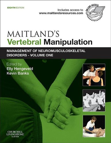
Maitland's vertebral manipulation
2014 · PDF

Maitland's vertebral manipulation
2014 · PDF

Maitland's Peripheral Manipulation
2013 · PDF